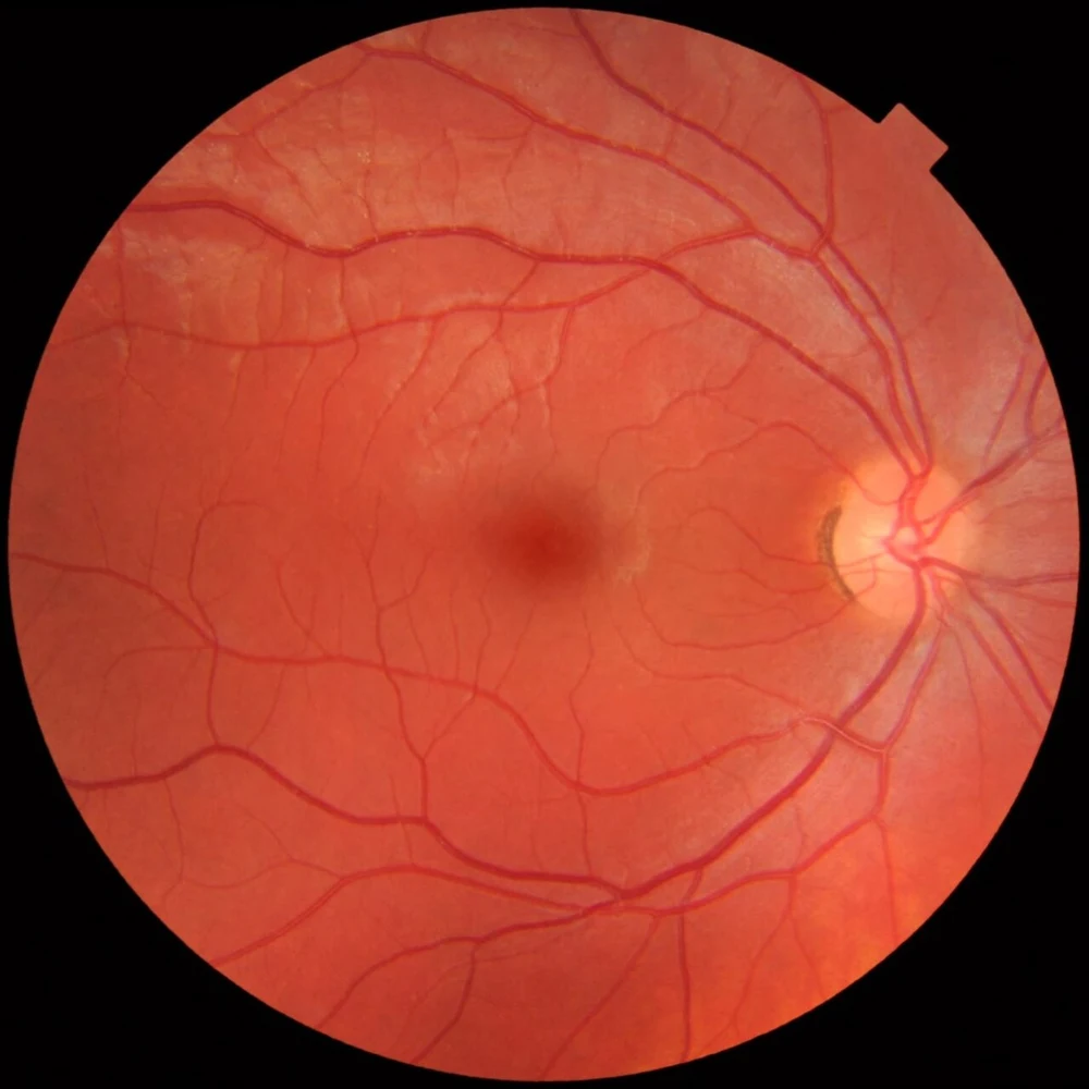
Retinal photography documents the retina, macula, vessels, and optic nerve. Comparing photographs over time can help identify change and communicate findings to another clinician.
A fundus photograph shows the retina from the front. This normal educational example from the credited independent source helps locate the vessels and optic nerve.Mikael Häggström · CC0 · Resized and converted to WebP; image content unaltered.
Photographs and scans answer different questions
Fundus and wide-field photographs show surface appearance. OCT provides cross-sectional measurements of tissue layers. We choose tests according to the clinical question; no image detects every disease or replaces the full examination.
When imaging may help
Imaging can support monitoring of diabetic retinopathy, macular degeneration, and glaucoma. Medication-related retinal screening uses specific testing protocols; a routine fundus photograph alone is not sufficient for every screening need.
Dilation and practical details
Imaging does not automatically replace dilation. Small pupils, cloudy media, artifacts, or a condition outside the photographed area can limit results. We discuss which tests are useful and any expected charges; coverage depends on the indication and benefits.
Stellest lenses combine a central prescription zone with surrounding lenslets designed to slow myopia progression. They are Midtown’s primary option to discuss for…
In July 2025 our Midtown Optometry team took our passion for eye health outdoors. Instead of spending the morning behind phoropters and slit‑lamps, we donned gloves and reusable b
Use readable text, reduce reflections, and place screens at a comfortable distance. Take breaks that fit your work, blink fully, and keep vents from blowing toward your…
Myopia often increases as a child’s eye grows. Specialized spectacle lenses, selected contact lenses, and 0.05% atropine can slow progression in appropriate patients.…
Dry eye can involve too little watery tear production, excessive evaporation, inflammation, or a combination of problems. Burning, grittiness, fluctuating blur, and…
On September 1, 2022, Midtown Optometry will be offering a free community clinic during regular business hours. We strive to provide high quality eyecare to everybody in…
Glaucoma is a group of diseases that damage the optic nerve. Eye pressure is an important risk factor and treatment target, but glaucoma can occur at pressures within the…
Presbyopia is the age-related loss of the eye’s ability to focus up close. It commonly becomes noticeable in midlife, but the timing and the correction that helps are…
You may have noticed that Midtown Optometry was shut down this Tuesday morning. The entire staff was out at Twin Lakes Beach doing a trash pick day. It is one of our favorite thing
AREDS-type supplements are for selected people with intermediate age-related macular degeneration, or late AMD in one eye, to lower progression risk in an eligible eye.…
Color vision deficiency means some colors are harder to distinguish. Most affected people do not see only in black and white. Inherited differences are often recognized in…
Children’s glasses should fit comfortably, support the prescribed vision correction, and suit the activities they actually do. Include your child in choosing a frame from…
Every sport has its own unique visual demands with regards to both vision and eye protection. Without the right pair of sports sunglasses or goggles, you could adversely…
While named after Frank Lloyd Wright, the complex at 550 Water Street was, in fact, designed Mr. Wright’s by his protege, Aaron Green in 1962. The complex is home to 400,000 square
Ultraviolet radiation is different from visible blue light. Excess UV exposure can injure the eye surface; long-term exposure is associated with some cataracts and other…
Conjunctivitis is inflammation of the thin tissue covering the white of the eye and inner eyelids. Viral and bacterial forms may spread between people; allergic…
Redness can come from dryness, allergy, infection, inflammation, injury, or a pressure problem. Appearance alone is not enough to choose treatment. Pain, light…
Do you know your dominant eye? Is it even possible to have a dominant eye? We are sure you have heard the term ‘dominant eye’ but you might not be sure what it means.…
Children use vision for many classroom and play activities, but there is no reliable universal percentage of learning that is “visual.” Learning difficulty has many…
We ask when symptoms began, whether one or both eyes are affected, about recent illness or exposure, and whether you wear contact lenses. The examination checks the cornea…