

October 22, 2026
Comparing Digital Lenses: Midtown, Costco & Warby Parker
A fair comparison of digital lens designs, fitting measurements, coatings, materials, and follow-up when choosing glasses in Santa Cruz.
Read more
A Closer Look
CIRRUS OCT, retinal and anterior-segment photos, IOLMaster biometry, Goldmann and iCare pressure checks, gonioscopy, and visual fields at Midtown in Santa Cruz.
Editorial update: October 22, 2026 · Midtown Optometry
Different tests answer different questions. At Midtown, we combine an eye examination with imaging and measurements when they are useful for your care. Your doctor explains what each test adds and how the results affect the plan.
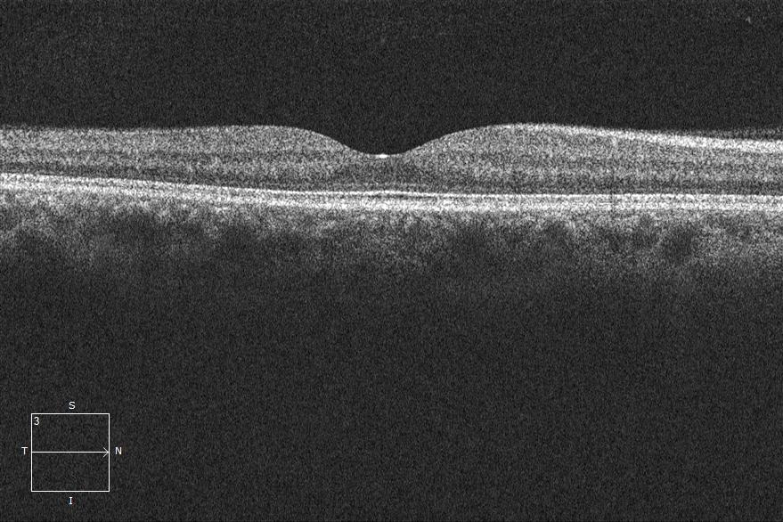
Optical coherence tomography uses light to build cross-sectional views of the retina and optic-nerve-related structures. It helps us monitor conditions such as glaucoma, macular degeneration, and diabetic retinal changes. Comparing scans over time can reveal changes that deserve closer attention.
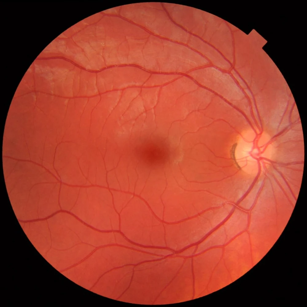
CLARUS captures wide-field retinal images. Fundus photographs document the appearance of the retina and optic nerve so we can compare visits and share findings when referring you. Photography and OCT provide complementary information. Imaging does not automatically replace dilation when a dilated examination is indicated.
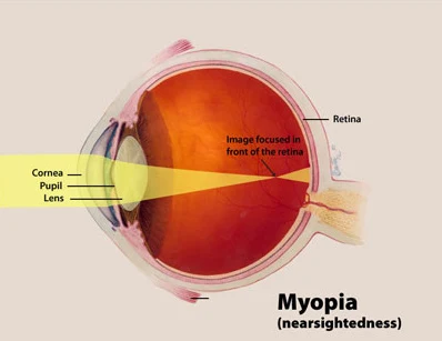
Our ZEISS IOLMaster optical biometer measures axial length—the distance from the front to the back of the eye. In myopia management, repeated measurements help us assess eye growth alongside changes in prescription. We use the trend, the child’s age, and the examination to guide decisions.
Our Radius headset-based visual-field system checks how well you detect lights in different areas of your vision. This provides functional information that complements OCT’s structural measurements, particularly in glaucoma care. Reliable testing sometimes requires repetition or confirmation with additional methods.
We also use OCULUS visual-field testing and Humphrey FDT, which uses frequency-doubling stimuli. These systems provide different testing approaches. Your doctor chooses a protocol and compares reliable results over time; values from different instruments need appropriate interpretation.
We use both Goldmann applanation and iCare rebound tonometry to measure eye pressure. Goldmann uses a small contact tip after numbing drops, usually with fluorescein dye. iCare uses a light, brief-contact rebound probe and generally needs no numbing drops. Technique and corneal properties influence measurements; we interpret the result alongside the examination and prior readings. iCare’s rebound technology.
With a specialized contact lens at the slit lamp, gonioscopy lets us inspect the angle where fluid drains from the eye. This helps distinguish open from narrow or closed angles and informs treatment and referral decisions, including whether a patient is suitable for SLT. Eye pressure, angle findings, optic-nerve structure, and visual function all contribute to glaucoma assessment.
At our examinations, we recommend retinal photographs, a screening five-line raster OCT, and anterior-segment photographs to create a record that can be compared at later visits. Retinal photos show the appearance of the back of the eye; the OCT samples cross-sectional retinal detail; anterior-segment photos document visible structures at the front of the eye. Together with the clinical examination, they help us recognize and follow changes.
The screening raster covers a limited retinal area. Symptoms or findings may call for dilation, a fuller macular scan, optic-nerve or nerve-fiber analysis, visual fields, or specialist assessment. Your doctor interprets the images in context and recommends the appropriate testing. Explore our imaging and examination tools.
Your findings and care plan guide the tests selected for each visit. The examination, symptoms, medical history, and follow-up plan determine what is appropriate. We discuss the reason for testing and any anticipated charges.
Original IOLMaster repeatability study in myopic children · ZEISS optical biometry.
PRACTICAL ANSWERS
Midtown uses CIRRUS OCT, CLARUS and fundus photography, anterior-segment imaging, ZEISS IOLMaster biometry, Goldmann and iCare tonometry, gonioscopy, and Radius, OCULUS, and Humphrey FDT visual-field testing. Your findings guide which tests are appropriate.
A retinal photograph records the appearance of the back of the eye. OCT measures cross-sectional retinal and optic-nerve structures. The images provide complementary information that your doctor interprets alongside the examination and other testing.
Axial length measures the eye’s front-to-back length. It adds information about eye growth that the prescription alone does not provide, helping us follow myopia progression over time.
How we monitor childhood myopia →We recommend retinal photographs, a screening five-line raster OCT, and anterior-segment images to establish a baseline and compare changes over time. Screening scans sample a limited area; findings can call for dilation or targeted diagnostic testing.
These educational resources support a conversation with your eye doctor. Your examination and medical history guide individual treatment decisions.
MIDTOWN OPTOMETRY · SANTA CRUZ