

October 22, 2026
Comparing Digital Lenses: Midtown, Costco & Warby Parker
A fair comparison of digital lens designs, fitting measurements, coatings, materials, and follow-up when choosing glasses in Santa Cruz.
Read more
Glaucoma Management
New glaucoma patients welcome in Santa Cruz: pressure checks, OCT, visual fields, medication management, and coordinated SLT referrals at Midtown Optometry.
Editorial update: October 22, 2026 · Midtown Optometry
Glaucoma care protects the optic nerve by detecting change and lowering eye pressure when needed. Midtown Optometry welcomes new glaucoma patients for evaluation and ongoing management in Santa Cruz, combining pressure measurements, optic-nerve imaging, and visual-field testing with a coordinated referral plan when laser or surgery is appropriate.
Bring your current eye-drop names and doses, previous OCT scans and visual-field results if available, and a history of laser or eye surgery. With your permission, we can request records to compare changes over time. Continue prescribed treatment unless your treating clinician changes the plan.
Open-angle glaucoma often has no early symptoms. It can damage the optic nerve and gradually reduce the visual field. Eye pressure is an important risk factor, but a single normal pressure reading does not rule out glaucoma, and elevated pressure does not always mean damage is present. Lost vision generally cannot be restored, so monitoring and treatment focus on preserving remaining sight.
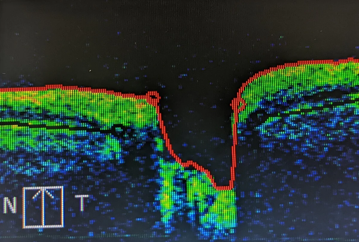
Diagnosis combines the examination with scan and field-test results. Your doctor looks for consistent patterns and may repeat testing to distinguish real change from measurement variability. Explore our diagnostic equipment.
We use both Goldmann applanation and iCare rebound tonometry to measure eye pressure. Goldmann uses a small contact tip after numbing drops, usually with fluorescein dye. iCare uses a light, brief-contact rebound probe and generally needs no numbing drops. Technique and corneal properties influence measurements; we interpret the result alongside the examination and prior readings. iCare’s rebound technology.
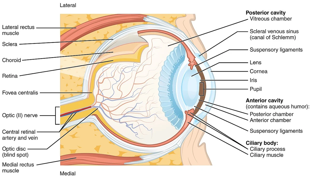
With a specialized contact lens at the slit lamp, gonioscopy lets us inspect the angle where fluid drains from the eye. This helps distinguish open from narrow or closed angles and informs treatment and referral decisions, including whether a patient is suitable for SLT. Eye pressure, angle findings, optic-nerve structure, and visual function all contribute to glaucoma assessment.
Our routine retinal photos, screening five-line raster OCT, and anterior-segment images establish useful baseline records. Glaucoma assessment also requires targeted optic-nerve and nerve-fiber testing and visual fields as indicated; a screening macular raster alone cannot rule out glaucoma.
An optometrist (OD) provides eye examinations, vision correction, and medical eye care within their training and licensure. An ophthalmologist (MD or DO) is a medical physician trained in eye care and surgery. An ophthalmologist’s degree is MD or DO; “OMD” is an informal label for the profession.
At Midtown, we manage appropriate glaucoma cases, prescribe and monitor treatment within our scope, and coordinate care with an ophthalmologist when laser, surgery, complex disease, or progression calls for specialist involvement. Dr. Fellers is glaucoma-certified. Referral adds another member to your care team, with follow-up shared according to your needs.
Why see an optometrist for glaucoma, and when might SLT be preferable to daily drops? Our article explains the roles of your care team and the research behind an SLT-first discussion.
SLT uses short laser pulses on the trabecular meshwork, part of the eye’s drainage system, to help fluid leave the eye more effectively and lower pressure. It is an outpatient procedure for suitable open-angle glaucoma or ocular hypertension. It can be offered as an initial treatment for suitable patients.
The ophthalmologist numbs the eye, places a special lens against it, and applies the laser to the drainage tissue. Follow-up checks pressure and response. Benefits can diminish, and repeat laser or additional treatment may be needed. Temporary discomfort, inflammation, blur, or a pressure rise can occur. SLT aims to protect remaining vision by lowering eye pressure. Existing glaucoma-related vision loss is permanent.
The original LiGHT trial randomized 718 people with newly diagnosed open-angle glaucoma or ocular hypertension to an SLT-first or drops-first pathway. At three years, 74.2% of patients in the SLT group needed no drops to maintain their target pressure. Read the 2019 Lancet trial.
At six years, 69.8% of SLT-first eyes reaching six years were at target pressure without glaucoma medication or trabeculectomy (a type of glaucoma surgery). Disease progression occurred in 19.6% of eyes in the SLT-first group versus 26.8% in the drops-first group. Some eyes also underwent cataract surgery. Repeat SLT was allowed, so the figures describe an SLT-first treatment pathway over six years. The later result reports eyes, while the three-year figure above reports patients. Read the six-year results and the 2025 correction with the updated tables.
We often recommend an SLT consultation with Mona Sane, MD, in Los Gatos when appropriate. Our team coordinates the referral, confirms the current office location, and shares the relevant testing. The treating ophthalmologist determines whether you are a good candidate and explains the procedure, risks, and follow-up.
The OHTS–EGPS model estimates five-year risk of early open-angle glaucoma in people with untreated ocular hypertension. Our educational points calculator uses five clinician-measured factors. Your doctor can help determine whether the model fits your situation.
This independent implementation of the published OHTS–EGPS simplified points model estimates the chance of developing early primary open-angle glaucoma in people with untreated ocular hypertension. Use measurements supplied by your eye-care clinician.
The model applies to people with elevated pressure and normal baseline discs and visual fields. Established glaucoma, treated pressure, visual disability, and individual treatment choices require a separate clinical assessment.
The pooled OHTS observation and EGPS placebo groups supplied the five predictors. Table 6 of the 2007 validation paper assigns each factor 0–4 points, then maps the total to one of five risk bands. The separate continuous model uses a different calculation and can give a different estimate.
The paper’s example totals 11 points, corresponding to 20%; its continuous estimate is 16.9%. Where published corneal-thickness bands overlap or leave a fractional boundary unspecified, this implementation directs you to the official continuous tool.
Your clinician considers additional findings, health, life expectancy, and preferences. A low score can coexist with eye disease that needs care. Continue your prescribed plan while reviewing results.
Primary validation study and points model · Washington University’s official calculators and limitations
Inputs stay in this page’s memory and clear when it reloads. This is Midtown’s educational implementation of the published model; Washington University’s software and PDFs remain on its website.
Different tools address different stages of care. De Moraes and colleagues’ 2012 visual-field progression model studied people with established, treated glaucoma: it was developed in 587 eyes and tested in an independent group of 62 eyes. It estimates the risk and rate of visual-field progression and requires clinical data and interpretation. Its purpose differs from predicting the first onset of glaucoma in ocular hypertension.
A 2023 OHTS medication-arm analysis explored risk estimation using pressure during treatment. It is a research extension; the calculator on this page retains the original untreated-pressure setting. For ongoing glaucoma care, we combine serial visual fields, OCT, pressure, and examination findings to assess change.
PRACTICAL ANSWERS
Yes. Pressure is one part of the assessment. Optic-nerve findings, visual fields, risk factors, and changes over time also matter.
Continued examinations and testing are necessary even when pressure responds well. Drops, repeat laser, or surgery may still be needed.
A suddenly painful red eye, halos with nausea, or sudden vision loss needs immediate assessment. Do not wait for a routine glaucoma appointment; seek emergency care if we cannot assess you promptly.
Midtown’s optometrists evaluate and monitor glaucoma, prescribe treatment within their scope, and coordinate ophthalmology referrals. Surgery and laser treatment are performed by the treating ophthalmologist.
Depending on the findings, we use eye-pressure measurements, optic-nerve examination, CIRRUS OCT, and Radius VR visual-field testing. The tests provide different information and are interpreted together over time.
Understand our diagnostic tests →We arrange SLT consultations and laser treatment with a referred ophthalmologist. We often refer to Dr. Mona Sane in Los Gatos and confirm the office when coordinating the referral.
Yes. We welcome patients who need glaucoma evaluation or ongoing management, including patients transferring care. Bring your current drops and previous testing if available. Advanced or progressing disease may also need ophthalmology involvement.
Request a glaucoma visit →For suitable open-angle glaucoma or ocular hypertension, SLT can lower pressure and reduce reliance on a daily drop routine. The LiGHT trial supports discussing it as an initial option. Suitability varies; some people need drops, repeat laser, or surgery.
Read our OD, ophthalmologist, and SLT guide →We use Goldmann applanation and iCare rebound tonometry, perform gonioscopy to inspect the drainage angle, and select visual-field testing from Radius, OCULUS, and Humphrey FDT systems. Results are interpreted with OCT, the optic-nerve examination, and prior records.
The model was developed for untreated ocular hypertension with normal baseline optic-nerve and visual-field findings. Published ranges include age 30–80, untreated Goldmann eye pressure 20–32 mmHg, and central corneal thickness 475–650 microns. It also uses vertical cup-to-disc ratio and standard automated visual-field pattern standard deviation. Your clinician should confirm its applicability.
OHTS estimates the chance of developing early glaucoma in ocular hypertension. Established glaucoma requires a separate assessment of pressure, optic-nerve structure, visual fields, and changes over time. Published progression models address that different question, with their own study populations and limitations.
These educational resources support a conversation with your eye doctor. Your examination and medical history guide individual treatment decisions.
MIDTOWN OPTOMETRY · SANTA CRUZ