

October 22, 2026
Comparing Digital Lenses: Midtown, Costco & Warby Parker
A fair comparison of digital lens designs, fitting measurements, coatings, materials, and follow-up when choosing glasses in Santa Cruz.
Read more
AMD / ARMD
AMD care in Santa Cruz: causes, smoking, heart health, diabetes, AREDS2 vitamins, current treatments, and emerging research with primary study links.
Editorial update: October 22, 2026 · Midtown Optometry
Age-related macular degeneration (AMD, also called ARMD) affects the retina’s central vision. Midtown Optometry evaluates and monitors AMD in Santa Cruz, explains supplement choices, and coordinates retina-specialist care. Treatment depends on the stage and whether there is leakage, atrophy, or both.
Research and U.S. treatment status checked October 22, 2026. Study links appear beside the claims they support; experimental treatments have their own section below.
New wavy lines, a central blurry or dark patch, or a sudden change in sight deserves prompt assessment. Call (831) 426-7172 today for guidance. Sudden substantial vision loss requires immediate eye care; our urgent-eye-care page explains how to seek help.
The macula is the small central region of the retina that helps you read, recognize faces, and see fine detail. AMD can affect these tasks while side vision remains useful. Early changes can be present before a person notices symptoms.
AMD develops through a combination of aging, inherited susceptibility, and environmental influences. Changes affect light-sensing cells, the retinal pigment epithelium that supports them, and nearby tissues. Oxidative stress, lipid handling, and inflammation contribute. Genetic studies have identified a role for the complement system, part of the immune response; this finding helped lead to treatments that target complement. NEI: the biology behind newer AMD treatments.
Yellowish deposits called drusen and pigment changes help an eye doctor assess stage and progression risk. Having drusen does not automatically mean severe vision loss is ahead. NEI: understanding AMD and its symptoms.
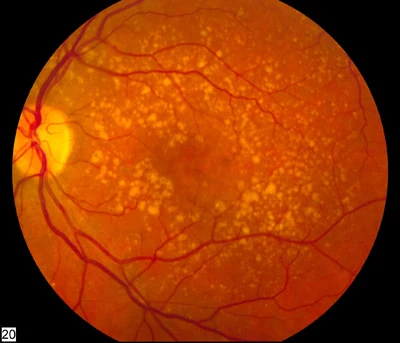
| Finding | What it means for care |
|---|---|
| Early dry AMD | Drusen or other early changes; monitoring and healthy habits are central to care. |
| Intermediate AMD | Larger drusen and/or qualifying pigment changes; discuss AREDS2 eligibility and individualized monitoring. |
| Geographic atrophy (GA) | An advanced dry form with areas of retinal cell loss; a retina specialist can assess treatment to slow enlargement. |
| Wet or neovascular AMD | Abnormal vessels leak fluid or blood beneath or within the retina; prompt retinal treatment can help preserve vision. |
Geographic atrophy and wet AMD can coexist. Examination and imaging identify the features that need treatment.
Increasing age and family history are important risk factors. Smoking is one of the strongest modifiable risks: it exposes retinal tissues to oxidative stress and is associated with progression to advanced AMD. Stopping smoking is a practical way to support both eye and general health. In the AREDS cohort, greater smoking exposure was associated with both neovascular AMD and central geographic atrophy. AREDS Report 19: progression risk factors.
AMD and cardiovascular disease share risk factors, including age and smoking. Studies also examine blood pressure, cholesterol, circulation, and inflammation. The association varies by population and AMD stage: the Cardiovascular Health Study found an association between early AMD and later coronary disease, while the ARIC study found no significant association for early AMD. These observational findings describe patterns; they cannot establish that one disease causes the other.
Keep blood pressure, cholesterol, activity, and cardiovascular care on track with your medical team. Your primary-care clinician assesses cardiovascular risk using your full health history. An AMD diagnosis alone does not diagnose heart disease.
Some studies associate diabetes with higher risk of advanced or wet AMD, and findings differ across studies and patient groups. In one higher-risk subgroup of AREDS, diabetes was associated with development of neovascular AMD. This supports discussing diabetes during an AMD assessment; it does not establish diabetes as the cause of AMD in an individual. Read the original AREDS risk-factor analysis.
Diabetic retinopathy is a separate condition caused by diabetes-related damage to retinal blood vessels. People can have both conditions. Glucose management and regular retinal examinations are especially important for diabetic eye health. Learn about diabetic eye care.
Make room for leafy green vegetables, a varied diet, regular physical activity that suits your health, and smoking cessation support when needed. Follow your clinician’s plan for blood pressure, cholesterol, and diabetes. These habits support overall health, and some are associated with lower AMD risk; they work alongside stage-specific eye care. NEI: lifestyle and inherited AMD risk.
Your eye doctor may recommend checking central vision in each eye separately, sometimes with an Amsler grid. Report a new change promptly, even between scheduled visits. Home checks complement retinal examinations and OCT. For established vision loss, better lighting, magnification, accessible device settings, and low-vision rehabilitation can make reading and everyday activities easier. NEI: low vision and rehabilitation.

AREDS2 is a targeted supplement for eligible AMD stages. The usual recommendation is for intermediate AMD in one or both eyes, or late AMD in one eye with an eligible fellow eye. Evidence supports reducing progression risk in these groups. For people with early AMD or healthy retinas, trials have not established a preventive benefit. Your examination determines eligibility. NEI: AREDS2 eligibility.
The original AREDS randomized trial, Report 8 (2001) established the benefit of antioxidants plus zinc in higher-risk participants. NEI summarizes the effect as roughly a 25% relative reduction in progression to advanced AMD over five years. The percentage describes a lower chance of progression; eyesight itself was a separate outcome. The result applies to the studied risk groups. NEI: interpreting the AREDS findings.
The AREDS2 randomized trial (2013) tested changes to that formula. Adding omega-3 DHA/EPA did not produce a significant additional reduction in advanced AMD in the primary analysis. The modern formula uses lutein and zeaxanthin in place of beta-carotene. AREDS2 Report 28 (2022), the long-term follow-up, supports that substitution and reinforces the lung-cancer concern with supplemental beta-carotene, especially for former smokers.
| Ingredient | Daily amount |
|---|---|
| Vitamin C | 500 mg |
| Vitamin E | 400 IU |
| Zinc | 80 mg, as zinc oxide |
| Copper | 2 mg, as cupric oxide |
| Lutein | 10 mg |
| Zeaxanthin | 2 mg |
Choose an AREDS2 formula without beta-carotene, particularly with a current or past smoking history. Copper helps offset zinc-related copper deficiency. Some products use 25 mg zinc; AREDS2 detected no significant difference between the zinc doses, and the original placebo-controlled benefit was established with 80 mg. Review the actual label and your medications with your clinician. These are high doses, and vitamin E and other ingredients can interact with medicines. NEI: formulation, zinc, and safety questions.
AREDS2 supports eligible patients alongside monitoring and treatment. Its goal is preserving future vision; it does not regrow lost retinal tissue. A 2024 secondary analysis of AREDS/AREDS2 found slower progression of noncentral geographic atrophy toward the fovea, the point of sharpest vision, in certain supplement comparisons. The overall atrophy-area growth comparisons were not significant. This post hoc finding adds a reason to discuss supplements with a retina specialist when atrophy is present; it is a different outcome from preventing advanced AMD.
Retina specialists use anti-VEGF treatment to suppress signals that drive abnormal blood-vessel growth and leakage. Eye injections can stabilize vision and sometimes improve it. The medication and interval depend on examination, OCT findings, response, and treatment history. Continued monitoring remains essential. Longer-lasting options can reduce injection frequency for some people: the TENAYA and LUCERNE randomized trials evaluated faricimab at intervals up to 16 weeks in selected patients, with similar average vision gains to the trial’s aflibercept regimen. Individual schedules vary.
Pegcetacoplan (Syfovre) and avacincaptad pegol (Izervay) are FDA-approved treatments for GA secondary to AMD. They target different complement proteins, C3 and C5, to slow enlargement of the atrophic area. A retina specialist weighs the location and growth of damage, useful vision, treatment burden, and risks. See the FDA Syfovre prescribing information and FDA Izervay prescribing information.
In OAKS and DERBY, pegcetacoplan slowed GA-area growth by 16–22% at 24 months, depending on the trial and dosing interval. The key visual-function outcomes did not show significant treatment-group differences at that time. In GATHER2, avacincaptad pegol reduced the rate of square-root-transformed GA-area growth by 14% over 12 months. These trials measured different populations and time periods, so their percentages do not establish which drug is better. Slower damage growth and better reading vision are separate outcomes.
Potential harms include eye infection, retinal detachment, increased eye pressure, and development of wet AMD. Syfovre’s labeling also warns of retinal vasculitis or vascular occlusion, which can cause severe vision loss. Your retinal specialist explains the expected benefit and the risks for your particular eye. These injections preserve tissue over time; they do not replace cells already lost.
The Valeda Light Delivery System uses controlled wavelengths of light, called photobiomodulation. FDA authorization covers a defined dry-AMD population with best-corrected vision of 20/32–20/70, qualifying drusen or noncentral atrophy, and no neovascular disease or center-involving GA. The FDA review describes an average benefit of about one eye-chart line compared with control after roughly two years. FDA De Novo review and eligibility.
LIGHTSITE III’s 24-month publication (2026) reports on 100 participants and 148 eyes receiving repeated treatment courses or sham treatment. The study was manufacturer-sponsored; broader replication and longer follow-up will help clarify durability and which patients benefit most. This is a prescription device with specific screening requirements, including photosensitivity considerations. Consumer red-light devices have different specifications and evidence. A specialist can discuss availability and suitability; this guide describes the research without listing Valeda as a Midtown service.
The following approaches remain investigational for AMD in the sources checked on October 22, 2026. Trial registration describes a research program; it does not establish effectiveness or FDA approval. A sponsor’s announcement is identified as such. Your retina specialist can help assess a trial’s eligibility, locations, potential benefit, and risks.
Surabgene lomparvovec (ABBV-RGX-314) delivers genetic instructions intended to let cells make an anti-VEGF protein over time. The aim is to reduce repeated injections while controlling leakage. A published phase 1/2a study followed 42 treated participants and supported further study of treatment durability. It was a small, open-label, dose-ranging trial. One high-dose participant had severe vision loss associated with pigmentary changes considered possibly treatment-related.
The larger randomized ATMOSPHERE phase 2/3 study and ASCENT phase 3 study compare subretinal gene therapy with standard anti-VEGF regimens. Their checked registry records contained no posted results. The sponsor’s August 2026 update projected pivotal results later in 2026. That is a company forecast; treatment availability depends on the results and regulatory review. Longer-term safety, durability, and any need for additional injections remain important questions.
AXPAXLI (OTX-TKI) is an investigational, dissolving axitinib implant intended to provide sustained suppression of blood-vessel growth signals. The SOL-1 phase 3 trial record identifies the study design. In a February 2026 sponsor announcement, the study met its vision-maintenance endpoint.
The comparison was a single implant versus a single aflibercept injection after initial loading, with rescue treatment available. That design tests durability under a particular protocol; it does not establish superiority over a regularly repeated aflibercept schedule. The sponsor’s second-quarter 2026 regulatory filing still described AXPAXLI as investigational. Detailed peer-reviewed results, repeated-dose safety, and regulatory decisions are key next milestones.
In a September 15, 2026 update, the sponsor continued to describe AXPAXLI as investigational and anticipated an FDA application in the fourth quarter of 2026. That is a company forecast; approval and availability depend on regulatory review.
OpRegen uses stem-cell-derived retinal pigment epithelial cells placed beneath the retina. The goal is to support surviving retinal cells in areas affected by GA. The GAlette phase 2a registry and Roche/Genentech’s trial description both describe an open-label, single-arm study of surgical delivery, safety, and preliminary activity.
The checked registry listed recruitment and no posted results. Without a randomized comparison, early improvements are difficult to separate from other influences. Larger controlled studies and long-term monitoring are needed to establish benefit and risks, including those of surgery and transplanted cells. A future cell-based treatment would need to demonstrate useful vision outcomes as well as survival of the implanted cells.
The research goals are fewer treatment visits for wet AMD, better ways to slow dry-AMD damage, and eventually support or replace damaged retinal tissue. Progress will likely involve different options for different stages. Approval dates and personal visual outcomes remain uncertain. Continue your current care plan while discussing new evidence or trial opportunities with your clinician.
Midtown recommends retinal photographs, a screening five-line raster OCT, and anterior-segment photographs during examinations as a baseline for future comparison. For suspected or established AMD, we select fuller macular OCT and other evaluation according to findings; a normal limited screening raster cannot exclude all macular disease.
We review your symptoms, family history, smoking history, medications, and general health. A retinal examination, CIRRUS OCT, CLARUS imaging, and fundus photography help us assess the macula and compare changes over time when indicated. OCT shows retinal layers and can help detect fluid; photographs document the retina’s appearance.
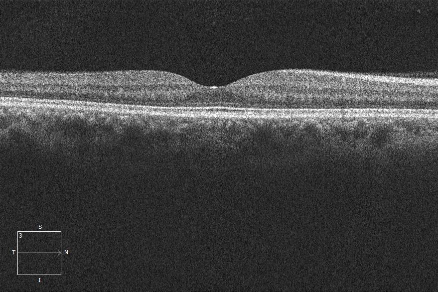
We discuss supplement suitability, home monitoring, follow-up intervals, and referral when retinal treatment is indicated. Retina specialists perform injections and other specialist procedures. Bring previous scans, injection records, and your supplement labels if you are transferring care. Request an AMD evaluation or use our first-visit checklist.
We accept VSP and Medicare. Benefits depend on the visit and service; our team can help clarify coverage.
Links above identify the original AREDS/AREDS2 trials, randomized treatment studies, FDA documents, trial registrations, and sponsor reports. Pay attention to the AMD stage, follow-up duration, and whether a result measures vision, damaged area, or injection frequency. Statistical averages describe study groups; your examination guides individual decisions.
For more context, read our eye-vitamin article, medical eye-care overview, and patient resources and evidence policy.
PRACTICAL ANSWERS
Both abbreviations mean age-related macular degeneration, a condition affecting the macula and central vision. Examination and imaging determine the stage and type.
Understand AMD stages →Smoking is a well-established modifiable AMD risk factor. Stopping smoking supports eye and cardiovascular health. Tell your eye doctor about current or past smoking, especially when discussing supplements.
AMD shares some risk factors with cardiovascular disease, and observational studies have found associations with cardiovascular disease or diabetes in certain groups. Results vary, and an association alone cannot establish cause. Diabetic retinopathy is a separate condition that can coexist with AMD.
Read the risk-factor evidence →AREDS2 is generally considered for intermediate AMD or an eligible fellow eye when one eye has late AMD. Your clinician should assess the stage, supplement label, medications, and health history. Newer secondary evidence about noncentral geographic atrophy also deserves an individualized discussion.
AREDS2 eligibility and research →The standard formula contains vitamin C, vitamin E, zinc, copper, lutein, and zeaxanthin. Adding DHA/EPA fish oil did not significantly reduce AMD progression in the AREDS2 primary analysis.
Care depends on stage. AREDS2 may help eligible patients. FDA-approved complement-inhibitor injections can slow geographic atrophy enlargement, and prescription Valeda light therapy is authorized for a specific dry-AMD population. A retina specialist assesses suitability, risks, and availability.
Current treatment options →The gene-therapy, axitinib-implant, and retinal-cell approaches discussed here remain investigational for AMD in the sources checked October 22, 2026. Trial eligibility and availability change. Ask a retina specialist about current studies and continue your prescribed care.
Explore the dated research update →Midtown evaluates and monitors AMD, reviews supplement suitability, and coordinates retina-specialist referrals. Retinal injections and specialist procedures are performed by the treating ophthalmology team. Call (831) 426-7172 or request an appointment.
Request an appointment →Call promptly for new distortion, a new central blurry or dark area, or a sudden change in sight. Sudden substantial vision loss needs immediate eye care. Home vision checks complement scheduled retinal examinations.
Urgent eye-care guidance →These educational resources support a conversation with your eye doctor. Your examination and medical history guide individual treatment decisions.
MIDTOWN OPTOMETRY · SANTA CRUZ